
Research Inspired by ‘Water Bears’ Leads to Innovations in Medicine, Food Preservation and Blood Storage
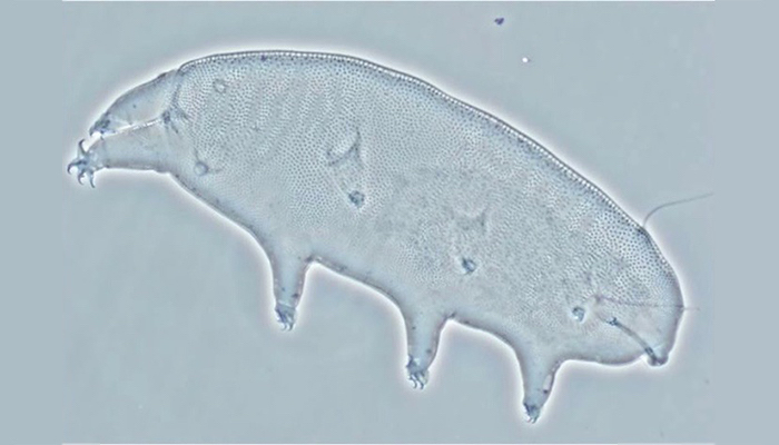
The study of microscopic tardigrades, also known as water bears, lead John Crowe and Lois Crowe to groundbreaking discoveries about the unique properties of a simple sugar known as trehalose. The Crowes’ method of preservation using trehalose allows the drug AmBisome® to be safely rehydrated after freeze drying. (Carl Johansson/UC Davis Bohart Museum)
When John Crowe and his wife Lois Crowe were researching tardigrades in the 1970s and 1980s, nobody knew much about how the speck-sized organisms — also known as water bears — were able to dry up completely, survive for years, and then somehow revive within a few hours when back in water.
Other organisms can do this as well. Brine shrimp, certain nematodes, baker’s yeast, and some desert plants can dry up for years and come back to life when there is water. The mechanism for this trick, though, was a mystery.
John was a professor in the UC Davis Department of Molecular and Cellular Biology and Lois wasa biophysicist in the UC Davis departments of Zoology and of Molecular and Cellular Biology. Together with their students and postdocs, they set out to discover how these organisms are able to survive in a desiccated form for years.
What they found is that nearly all these organisms produce a simple sugar known as trehalose. They also found that the organisms convert as much as 20 percent of their dry weight to trehalose before they can be dried.
The Crowes were able to show that trehalose acts as a water replacement — protecting cells by preventing cell membranes from falling apart and stabilizing proteins and nucleic acids in the dry state.
Their discoveries about the cell-protecting abilities of trehalose opened the door for a wide range of new innovations in food preservation, medicine and blood storage.
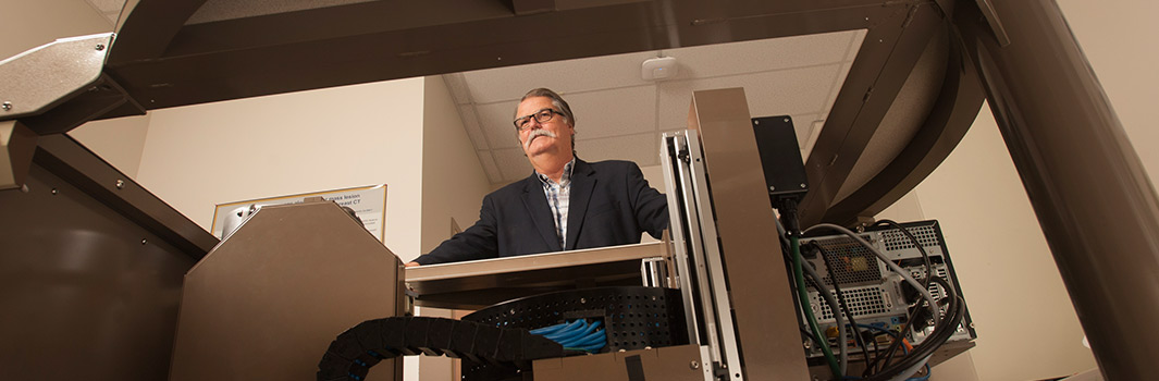
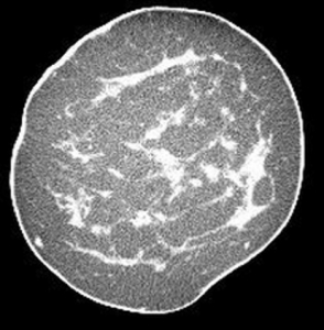 regular screening, but Boone has developed what could be a better approach, hopefully improving both detection and patient outcomes. Boone and his team have designed and developed an innovative computed tomography (CT) scanner designed specifically for imaging the breast (UC Case 2005-543). The intended advantage of this device is that it provides a true three-dimensional, highly-detailed image of the human breast, offering a less obstructed view of potential lesions than the current two-dimensional mammogram.