Posts

UC Davis Names Recipients of 2020 Chancellor’s Innovation Awards

Honoring Advances in Medical Imaging, Infant Health and Pain Relief, Plus Commitment to Building Aggie Square

Recipients of 2020 Chancellor’s Innovation Awards

Original post: ucdavis.edu/news/uc-davis-names-recipients-2020-chancellors-innovation-awards

The University of California, Davis, today (June 15) named the recipients of the 2020 Chancellor’s Innovation Awards. The awards recognize faculty, project teams and community partners for their work, dedication and success in improving the lives of others and addressing the needs of our global society through innovative solutions.

“Research universities like UC Davis play a critical role in advancing innovative solutions for the global community that not only stimulate our economy but create a better quality of life,” Chancellor Gary S. May said. “The recipients of this year’s awards demonstrate the impact of reaching beyond what is expected to deliver game-changing innovations that address some of the world’s most critical issues.”

The awards comprise Innovator of the Year, Innovative Community Partner and Lifetime Achievement in Innovation. The program is managed by the Office of Research.

“Some of the greatest examples of bold innovation emerge when experts from different disciplines work together to solve a problem,” said Prasant Mohapatra, vice chancellor for research. “Many of the recipients for this year’s awards illustrate just how effective those collaborations can be.”

Read more..

Startup Uses Advanced Imaging Technology and Machine Learning to Sort Seeds and Insects

UC Davis startup Spectral Analytix applies machine vision, robotics and machine learning to automatically classify or sort seeds and insects.

UC Davis startup Spectral Analytix applies machine vision, robotics and machine learning to automatically classify or sort seeds and insects. (Hector Amezcua/UC Davis)

Original Post: research.ucdavis.edu/startup-uses-advanced-imaging-technology-and-machine-learning-to-sort-seeds-and-insects/

Christian Nansen, an associate professor in the UC Davis Department of Entomology and Nematology, has launched a startup, Spectral Analytix, to apply machine vision and machine learning to the classification and sorting of seeds and insects.

“The idea is to combine machine vision, robotics and machine learning so you have an automatic eye, an automated arm and an automated brain,” said Nansen. “If you automate those three components you end up with a system that can automatically classify or sort whatever you are working with.”

For the machine “vision,” Nansen works with hyperspectral cameras, which collect data at very high spectral resolution. “The camera on your phone divides light into three wavelengths—red, blue and green,” said Nansen. “You can think of it like a cake with three layers—for each pixel you have three values. With a hyperspectral camera you have 250 bands, so the ‘cake’ now has 250 layers.”

Hyperspectral imaging is used for a wide variety of applications, from mining to surveillance to investigating works of art. Paired with machine learning, hyperspectral imaging is widely used in food processing and recycling industries for sorting.

Several aspects of crop breeding and commercialization of crop seeds involve inspection and quality control.

“Often, these inspection and control measures are time consuming and rely on highly trained technicians. They may also be associated with consistency challenges due to human error. So, replacing them with automated procedures can improve such inspection and control measures and also enable people to focus on other tasks that involve higher levels of complexity,” said Nansen.

Read more…


EXPLORER, a UC Davis imaging breakthrough, makes a media splash

(SACRAMENTO) — EXPLORER, the world’s first total-body positron emission tomography (PET) scanner that can capture a 3D picture of the whole human body at once, is up and running at UC Davis Health.

Developed by UC Davis scientists, EXPLORER has already captured the attention of radiology experts around the world. It was featured in an article in Nature and its images have drawn hundreds of thousands of views on YouTube. The scanner and its inventors were introduced to local media outlets on Monday.

Image quality a game changer

EXPLORER’s exceptional image quality gives it nearly limitless potential applications for both clinical use and research.

“We are thrilled, after almost 15 years, to finally have brought this concept of total-body imaging to fruition,” said co-inventor Simon Cherry, distinguished professor in the UC Davis Department of Biomedical Engineering. “The first images coming off the EXPLORER scanner have exceeded what we, and I think many others in our field, thought would be possible.”

The EXPLORER scanner, which combines PET and x-ray computed tomography (CT), was installed in May in a specially prepared space on Folsom Boulevard. Built by UC Davis industry partner United Imaging Healthcare (UIH), EXPLORER was shipped in two 40-foot containers to a warehouse in Oakland. From there, the parts arrived by truck in several deliveries.

Read more…

Explorer team selected by Physics World as one of the Top 10 Breakthroughs in 2018

Original post: bme.ucdavis.edu/news/explorer-included-breakthrough-year-physics-world

Editor Tami Freeman of Physics World chose the EXPLORER total-body PET system as one of her top-five “Breakthroughs of the Year,” and the publication’s entire editorial team named EXPLORER among its top-10 2018 breakthroughs:

“The EXPLORER PET/CT scanner – the world’s first medical imaging system that can capture a 3D image of the entire human body simultaneously – has produced its first human images. Developed by UC Davis scientists and a multi-institutional consortium, EXPLORER can scan up to 40 times faster, or use up to 40 times less radiation dose, than current PET systems, making it possible to conduct repeated studies in an individual, or dramatically reduce dose in paediatric studies. The high-sensitivity scanner can also create movies that track radiolabelled drugs as they move around the body.”

Editors based their annual choices on three criteria:

  • Significant advance in knowledge or understanding
  • Importance of work for scientific progress and/or development of real-world applications
  • Of general interest to Physics World readers.

Read Freeman’s article and the top-10 article in their entireties.

Improving detection of breast cancer

Improving detection of breast cancer

Dr. John Boone is a recognized expert in the field of medical imaging, with a focus on improving breast cancer detection. He and his team have developed a device with the potential to detect tumors in the breast earlier and with less discomfort.

The American Cancer Society reports that breast cancer is the second most common form of cancer among American women, following skin cancers. It estimates that about 1 in 8 women in the U.S. will develop invasive breast cancer during her lifetime.

Traditionally, mammograms have been used to detect breast cancers as part of aboonebreastct_300dpi-003 
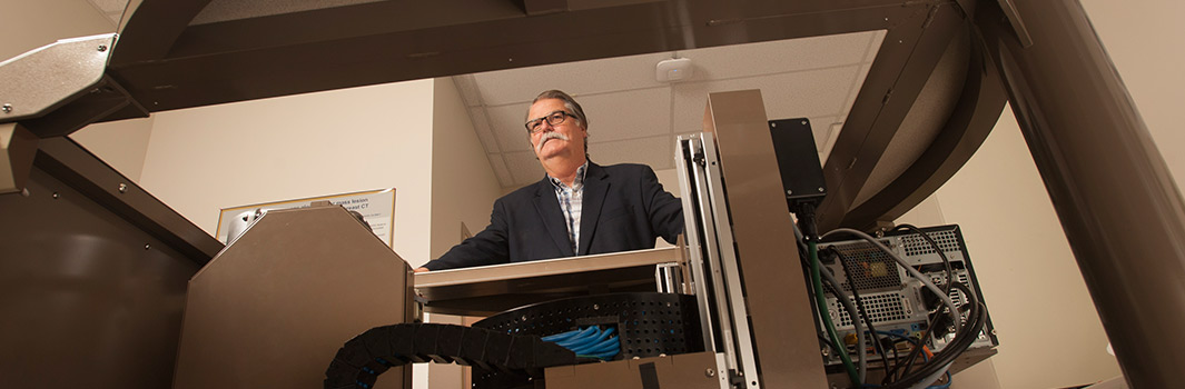
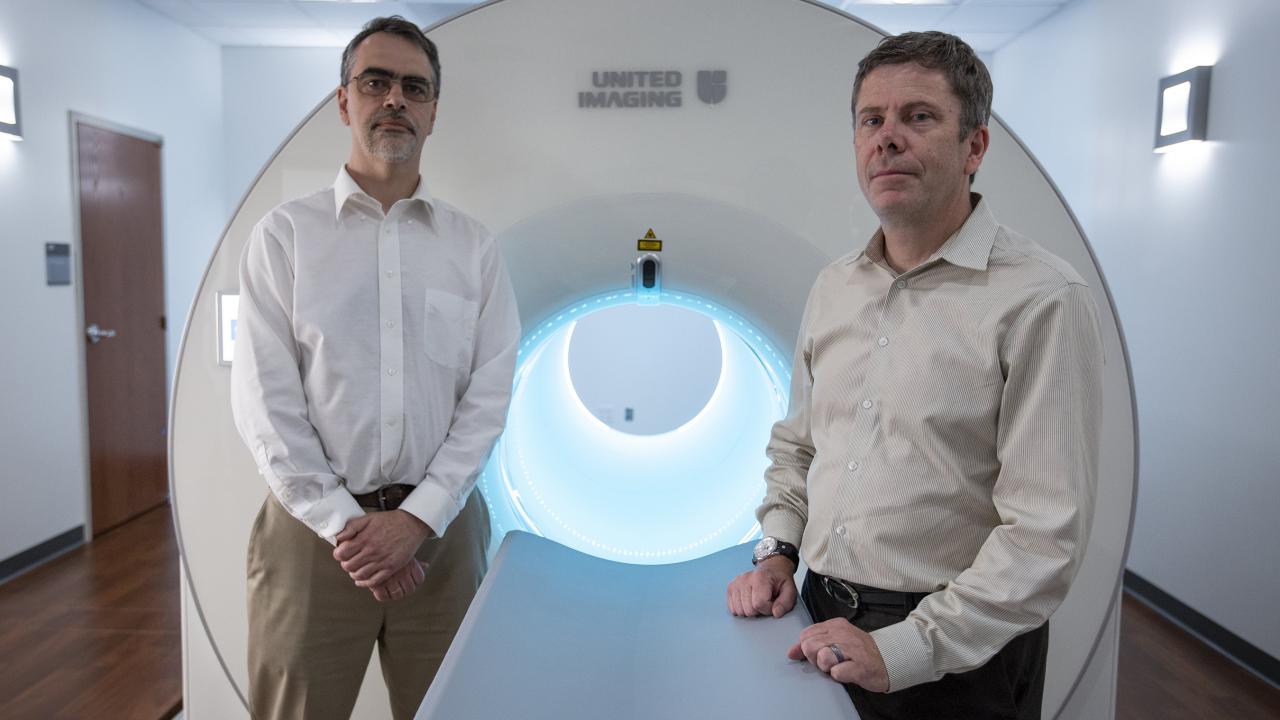
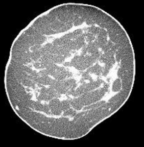
regular screening, but Boone has developed what could be a better approach, hopefully improving both detection and patient outcomes. Boone and his team have designed and developed an innovative computed tomography (CT) scanner designed specifically for imaging the breast (UC Case 2005-543). The intended advantage of this device is that it provides a true three-dimensional, highly-detailed image of the human breast, offering a less obstructed view of potential lesions than the current two-dimensional mammogram.

Unlike mammography, the scanner does not require compression of the breast. Instead, the patient lies face down on a padded table and places the breast in a circular opening. The scanner generates 300 to 500 tomographic image “slices” of the breast, which are then assembled into a three-dimensional digital model. The imaging procedure takes approximately 10 seconds.

Thanks to support from the National Institutes of Health, Boone’s team has assembled four scanners that have been used to image over 600 women at the UC Davis Medical Center.

The technology led to the formation of Isotropic Imaging Corporation, which intends to license the technology developed at UC Davis to commercialize a scanner.