EXPLORER, a UC Davis imaging breakthrough, makes a media splash
Developed by UC Davis scientists, EXPLORER has already captured the attention of radiology experts around the world. It was featured in an article in Nature and its images have drawn hundreds of thousands of views on YouTube. The scanner and its inventors were introduced to local media outlets on Monday.
Image quality a game changer
EXPLORER’s exceptional image quality gives it nearly limitless potential applications for both clinical use and research.
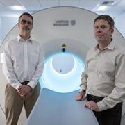
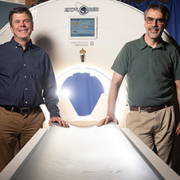
“We are thrilled, after almost 15 years, to finally have brought this concept of total-body imaging to fruition,” said co-inventor Simon Cherry, distinguished professor in the UC Davis Department of Biomedical Engineering. “The first images coming off the EXPLORER scanner have exceeded what we, and I think many others in our field, thought would be possible.”
The EXPLORER scanner, which combines PET and x-ray computed tomography (CT), was installed in May in a specially prepared space on Folsom Boulevard. Built by UC Davis industry partner United Imaging Healthcare (UIH), EXPLORER was shipped in two 40-foot containers to a warehouse in Oakland. From there, the parts arrived by truck in several deliveries.

